Nervous Anatomy Lessons

Human anatomy is the basis of surgical practice. The complexity and beauty of the human body is a joy to study. I hope to add more and more content here: If you find this useful or would like other specific content please leave a message below.

Brachial Plexus Anatomy

A graphic tour of some of the basic parts of brachial plexus anatomy

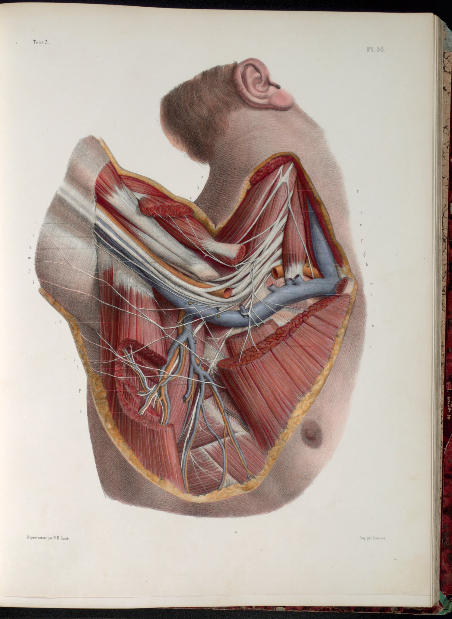
The brachial plexus is a wonderful part of the body, many colleagues ask me for teaching on this part of anatomy and some patients are interested in seeing and learning more about their bodies.

For many people, medical images can be considered ‘gory’. I think human anatomy is beautiful and think the democratisation of knowledge about our bodies is important. So I have published the guide below using a wonderful historic image (by Nicolas Henri Jacob an early 19th Century Painter).

If you do not want to see human anatomy please do not scroll any further: To return to the home page click here

C5 Nerve root: The nerve that comes from the spine at the level of Cervical level 5 (C5) controls the shoulder. It brings sensation back from the shoulder joint skin over the shoulder and muscles to move the shouder out and forward and back.
C6 Nerve root: The nerve that comes from the spine at the level of Cervical level 6 (C6) controls the elbow bending muscles and some of the wrist bending and pronating muscles. Sensation is to the outer upper arm and outer forearm down to the thumb and index finger.
C7 Nerve root: The nerve that comes from the spine at the level of Cervical level 7 (C7) is motor across a wide collection of muscles of the elbow, forearm and chest. The C7 sensory dermatome is to the middle finger.
Subclavian artery; this is the blood vessel that brings blood from the heart to the arm. It runs from the aorta on the left but as shown here but on the right it is often the first branch of the brachiocephalic trunk. It lies behind the scalenius anterior muscle and runs over the first rib behind this muscle’s insertion with the lower trunk and the rest of the plexus in the thoracic outlet.
Lower Trunk – is made by the lowest cervical root (C8) and the first thoracic root (T1). This trunk brings movement to the hand and feeling to the inner arm and little and ring fingers.
The phrenic nerve is the nerve from C3,4,5 which controls the diaphragm breathing muscle. This large muscle sits between the chest and the abdomen. In embrology the muscles that form the diaphragm started in the neck and migrated into the chest this is why it is controlled by the phrenic (a middle cervical nerve) and not via the chest intercostals, as the other muscles of breathing are.
The supracscapular nerve is commonly the first large branch of the upper trunk but often this can branch purely from C5 before the confluence of the upper trunk. This muscle is a pure motor nerve and controls the supraspinatus and infraspinatus muscles. These elevate, abduct, and externally rotate the glenohumeral joint.
Divisions of the upper trunk – the upper trunk (C5,6) splits in to anterior (ADUT) and posterior (PDUT) divisions of the upper trunk. ADUT leads to the lateral cord: lateral pectoral nerves musculocutaneous nerve and the lateral contribution to the median. PDUT runs to the posterior cord mainly to the axillary nerve but also upper parts of the radial nerve.
Posterior cord is the confulence of the posterior divisions of the upper middle and lower trunks. It brings elemetns of C5-T1 to the function of adduction, abduction, retroposition and internal rotation of the sholder, extension of shoulder, elbow, wrist and fingers. It gives off the upper and lower subscap nerves and thoracodorsal nerve (to Latissimus dorsi) to then terminate as the axillary and radial nerves.
Long thoracic nerve has root levels of C5,6,7. it runs through the scalenius medius to run behind this muscle over the first rib and down on to the chest wall to innervate the Serratus anterior muscle which stabilises and protracts the scapula.
Thoracodoral nerve has root levels C6,7,8 and is s pure motor nerve innervating the Latissimus dorsi muscle. This muscle adducts and internally rotates the shoudler. You can follow it here running with the thoracodorsal pedicle to the motor point within the muscle.
Subclavian vein. This is the point of drainage of venous blood from the arm to then run in to the right atrium via the superior vena cava.
Medial clavicle. This is the cut end of the collar bone (clavicle) in this picture a section has been taken away to show the anatomy. This is occasionally necesary in surgery to the brachial pleus. Normally all of the structures of the brachial pleuxus run under the structure in an area of the body called the thoracic outlet.
Thoracodorsal pedicle. The branches of the artery and vein to run with the nerve and supply the Latissiumus dorsi muscle. This long pedicle allows this muscle to be mobilised as a pedicled transfer or a free graft.
Lateral contribution to the Median nerve (LCMN) – this is the C5,6,7, contribution to the median nerve from the lateral cord. These nerves supply the sensory fibres to the median nerve and motor to the proximal muscles- pronator, flexor carpi radialis, palmaris longus mainly.
Medial contribution to the Median nerve (MCMN) – this is the C8,T1, contribution to the median nerve from the medial cord. These nerves supply no sensory fibres to the median nerve and motor to the extrinsic and intrinsic flexors of the fingers and hand.
Median nerve- this is the median nerve proper lying ventral to the brachial artery and formed by the LCMN and MCMN. In brachial plexus dissection this is the middle limb of the ‘M’ which is first found then can be followed to the musculocutanoneous nerve via the LCMN and to the Ulnar nerve by the MCMN.
Musculocutaneous nerve (a branch of the lateral cord) flexes the elbow and bring sensation from the lateral cutaneous nerve of the forearm.
Ulnar nerve this branch of the medial cord brings function to the long flexors and sensation to the little and ring fingers as well as the ulnar intrinsic muscles.